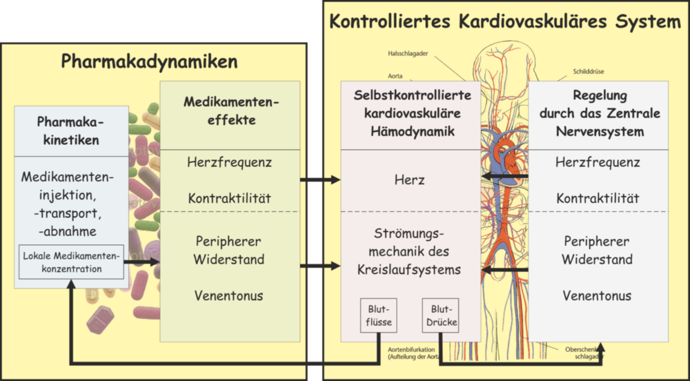
Objective
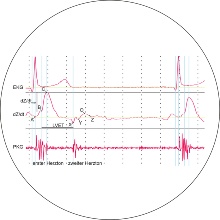
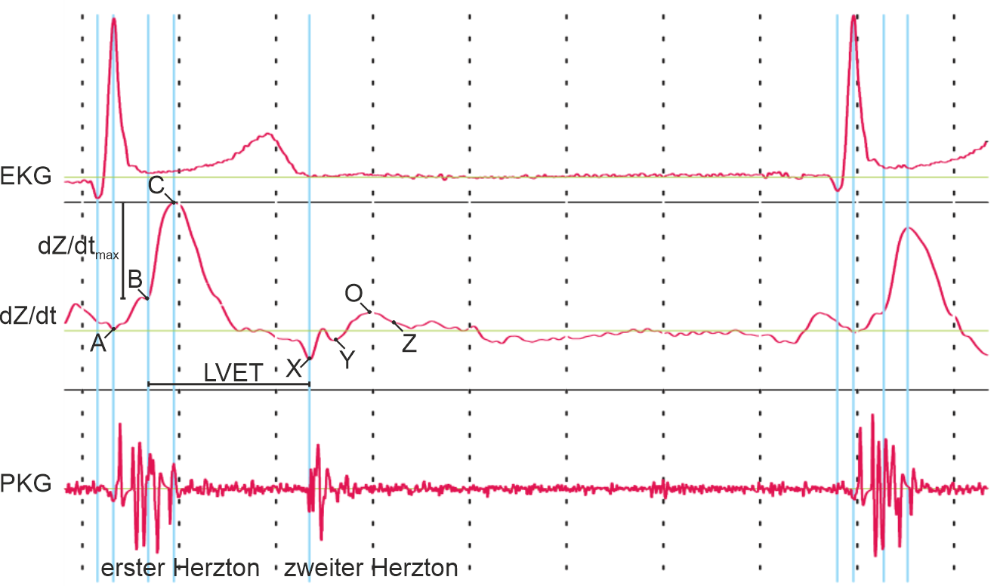
Impedance plethysmography (IPG) is a non-invasive method for measuring the electrical resistance of the subcutaneous tissue. With impedance cardiography (ICG) as a special form of IPG, the time-dependent thoracic resistance is measured to estimate hemodynamic parameters such as the stroke volume or the contractility of the heart. However, the determination of absolute stroke volume values is often unreliable and hardly reproducible. This is mainly, because the values are determined from a 1D measurement of the electrical thoracic impedance. At BMT, we investigate alternative forms of ICG basing on multidimensional impedance measurements for a highly sensitive stroke volume estimation in cardiac diagnostic.
Projects
Development
We developed both an IPG-system for the simultaneous mesasurement of three independent impedance signals and a hardware simulator for its calibration. The interface to the computer is provided by multifunctional input/output devices from National Instruments. The acquisition of the data as well as their graphical visualization and analysis are performed by means of a self-developed software program based on a LabVIEW-platform (National Instruments). In additon, we implemented a software compartment model of the cardiovascular system using Simulink (MathWorks). This model allows us to investigate suitable electrode configurations for the impedance measurement on different (orthogonal) trajectories and finally to reconstruct the measured signals to identify their origins.
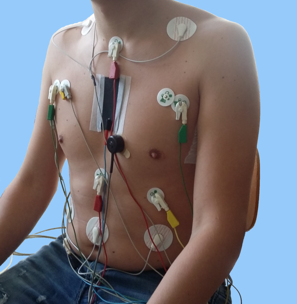
Investigation
The most challenging tasks are to find the best positions for the electrodes for measuring three orthogonal IPGs, and to derive a suitable alogrithm, with which the stroke volume can be determined reliably and reproducibly. For this purpose, we investigate in experiments different electrode configurations and electrode materials on phantoms and subjects in combination with our software model. We use the obtained data for the reconstruction of the measured signals with our software model in order to identify their sources. From the results we derive an algorithm to determine the stroke volume.
Contact

Johannes Port
Dr.-Ing.Deputy Head

Giorgio Cattaneo
Prof. Dr.-Ing.Head of the institute, Academic dean Master Medical Engineering